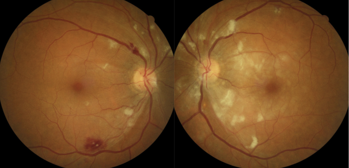

A 2010 population-based study indicated four novel loci (19q13, 6q24, 12q24, and 5q14) as significantly associated with retinal venular caliber. Some studies suggest a genetic influence on retinal vascular caliber. The major risk factor for malignant hypertension is the degree of blood pressure elevation over normal. The major risk for arteriosclerotic hypertensive retinopathy is the duration of elevated blood pressure. Risk factors for essential hypertension include high salt diet, obesity, tobacco use, alcohol, family history, stress, and ethnic background. In addition, genetic factors have been found to be associated with a higher risk of hypertensive retinopathy. Many young patients with secondary hypertension may actually present to an ophthalmologist with bilateral vision loss due to serous macular detachment, bilateral optic disc edema, and exudative retinal detachment. However, secondary hypertension can develop in the setting of pheochromocytoma, primary hyperaldosteronism, Cushing’s syndrome, renal parenchymal disease, renal vascular disease, coarctation of the aorta, obstructive sleep apnea, hyperparathyroidism, and hyperthyroidism. Essential hypertension is a polygenic disease with multiple modifiable environmental factors contributing to the disease. Primary hypertension is usually essential and not secondary to another disease process. The arteriosclerotic changes of hypertensive retinopathy are caused by chronically elevated blood pressure, defined as SBP greater than 140 mmHg and DBP greater than 90 mmHg. Men are more affected than women in age groups less than 45 years old and women are more affected in age groups greater than 65 years old. In addition, the incidence of blood pressure increases with age. Hypertensive retinopathy is more common in African Americans and Chinese descent. Hypertensive retinopathy ranges from 2-17% in non-diabetic patients but the prevalence varies by demographic groups. In the United States, 33% of adults have hypertension and only 52% have controlled blood pressures. The chronic effects of hypertension are caused by arteriosclerosis and predispose patients to visual loss from complications of vascular occlusions or macroaneurysms. The acute effects of systemic arterial hypertension are a result of vasospasm to autoregulate perfusion. Hypertensive retinopathy includes two disease processes. The American College of Cardiology/American Heart Association (ACC/AHA) suggested the following definitions for high blood pressure in 2017.

The arteriosclerotic changes of hypertensive retinopathy are caused by chronically elevated blood pressure. This article focuses primarily upon hypertensive retinopathy, which is the most common ocular presentation.

Moreover, hypertension can also cause occlusion of major retinal vessels such as the branch retinal artery, central retinal artery, branch retinal vein and central retinal vein. Specifically, hypertension may lead to multiple adverse effects to the eye that can inevitably cause cause retinopathy, optic neuropathy, and choroidopathy. Hypertension is a risk factor for systemic conditions that can lead to target-organ damage.


 0 kommentar(er)
